Table of Contents
Unraveling the Mysteries of Multifocal Pigmentation
Intro
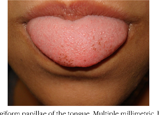
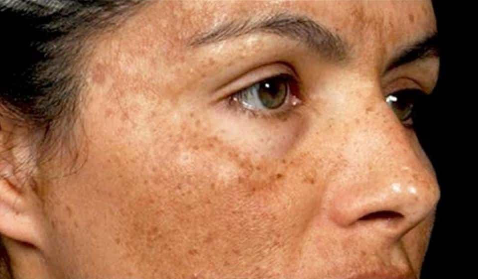
When it comes to understanding the mysteries of Multifocal Pigmentation, it helps to first consider the two different types of pigmentation: diffuse and multifocal. Diffuse pigmentation refers to general pigmentation that is caused by a variety of factors, such as drugs, systemic diseases, and environmental toxins. Multifocal Pigmentation, on the other hand, is a more focused form of pigmentation that can include amalgam tattoos, oral melanotic macules, melanocytic nevi, and melanoacanthoma.
Understanding Diffuse Pigmentation
Diffuse pigmentation is a broad term that encompasses various forms of pigmentation disorders. It refers to the general pigmentation that is caused by a range of factors, including drugs, systemic diseases, and environmental toxins. Unlike multifocal pigmentation, which is a more focused form of pigmentation, diffuse pigmentation affects a larger area and can occur in different parts of the body.
Physiological pigmentation is one type of diffuse pigmentation that occurs naturally in certain individuals. It is typically more common in people with darker skin tones. This type of pigmentation can be influenced by factors such as hormones, sun exposure, and genetic predisposition.
Drug-induced melanosis is another form of diffuse pigmentation that occurs as a side effect of certain medications. These medications can include antibiotics, antimalarials, chemotherapeutic agents, and psychotropic drugs. The pigmentation can manifest in various areas of the body, such as the face, hands, or nails.
Melanosis associated with systemic diseases refers to pigmentation changes that occur as a result of underlying medical conditions. Conditions such as Addison’s disease, hemochromatosis, and hyperthyroidism can lead to diffuse pigmentation.
Smoker’s melanosis is a specific type of diffuse pigmentation that affects individuals who smoke. It is characterized by dark pigmentation in the oral cavity, including the gums, lips, and buccal mucosa.
Heavy metal pigmentation occurs when metals such as lead, arsenic, or mercury accumulate in the body and cause changes in skin pigmentation. Occupational exposure, contaminated food or water, and certain medications can contribute to heavy metal pigmentation.
In summary, diffuse pigmentation encompasses various types of pigmentation disorders, including physiological pigmentation, drug-induced melanosis, melanosis associated with systemic diseases, smoker’s melanosis, and heavy metal pigmentation. Understanding these different types can help in identifying and managing pigmentation issues effectively.

Different Types of Multifocal Pigmentation
Multifocal pigmentation is a more focused form of pigmentation that can present itself in various ways. Understanding the different types can help identify and manage pigmentation issues effectively.
1. Amalgam Tattoos: These are caused by the deposition of amalgam particles in the oral cavity. It usually occurs when amalgam fillings are placed, removed, or repaired. The pigmentation can range from gray to black and is typically found on the gums or other soft tissues.
2. Oral Melanotic Macule: This is a small, flat, brown spot that appears on the lips or oral mucosa. It is considered a benign lesion and is often seen in individuals with darker skin tones. Although it is usually harmless, it is important to have any new or changing pigmented lesions evaluated by a healthcare professional.
3. Melanocytic Nevi: Also known as moles, these are common pigmented growths on the skin. They can be flat or raised and may vary in color. While most moles are benign, some may have the potential to develop into skin cancer, so it is important to monitor them for any changes in size, shape, or color.
4. Melanoacanthoma: This is a rare benign pigmented lesion that typically occurs on the oral mucosa. It is characterized by a brown or black papule or plaque. Melanoacanthoma is typically asymptomatic and does not require treatment, but it is important to have it evaluated by a healthcare professional to rule out any potential concerns.
Causes and Symptoms of Multifocal Pigmentation
Multifocal pigmentation can have various causes, each with its own distinct symptoms. Understanding these causes and symptoms is crucial in effectively managing and treating pigmentation issues.
One common cause of multifocal pigmentation is amalgam tattoos. These tattoos occur when amalgam particles from dental fillings are deposited in the oral cavity. They often appear as gray or black spots on the gums or other soft tissues.
Another cause of multifocal pigmentation is oral melanotic macule. This is a benign lesion characterized by small, flat, brown spots that can appear on the lips or oral mucosa. While usually harmless, it is important to have any new or changing pigmented lesions evaluated by a healthcare professional.
Melanocytic nevi, also known as moles, can also contribute to multifocal pigmentation. These common pigmented growths can vary in color, shape, and size. While most moles are benign, some may have the potential to develop into skin cancer, so it is important to monitor them for any changes.
Lastly, melanoacanthoma is a rare benign pigmented lesion that typically occurs on the oral mucosa. It is characterized by brown or black papules or plaques.
In summary, multifocal pigmentation can be caused by amalgam tattoos, oral melanotic macules, melanocytic nevi, and melanoacanthoma. Recognizing these causes and their associated symptoms is essential in determining the appropriate course of action for managing and treating multifocal pigmentation.

Diagnosis and Treatment Options
Diagnosing multifocal pigmentation requires a comprehensive evaluation by a healthcare professional. They will examine the affected areas, inquire about the patient’s medical history, and may order additional tests or biopsies to determine the exact cause of the pigmentation.
For amalgam tattoos, a dental examination is typically conducted to identify the presence of amalgam particles in the oral cavity. X-rays or other imaging tests may be used to confirm the diagnosis.
Oral melanotic macules are usually diagnosed through a visual examination. In some cases, a biopsy may be performed to rule out any underlying malignancies.
Melanocytic nevi can be diagnosed through a physical examination, as well as dermoscopy or a skin biopsy if necessary. If a mole shows signs of atypical features or changes, further evaluation may be needed to assess the risk of skin cancer.
Melanoacanthoma is typically diagnosed through a clinical evaluation and a biopsy of the affected area to confirm the benign nature of the lesion.
Treatment options for multifocal pigmentation depend on the specific cause and severity of the pigmentation. In some cases, no treatment may be necessary, especially if the pigmentation is benign and not causing any symptoms. However, if the pigmentation is causing aesthetic or functional concerns, various treatment options may be considered.
For amalgam tattoos, professional dental cleaning or bleaching procedures may help lighten the pigmentation. In more severe cases, surgical excision or laser treatments may be recommended.
Oral melanotic macules usually do not require treatment unless they cause cosmetic concerns. In such cases, cryotherapy or laser therapy may be used to remove or lighten the pigmentation.
Melanocytic nevi are generally monitored for changes, and if any signs of malignancy are present, surgical removal may be recommended.
Prevention Measures and Lifestyle Changes
While it may not be possible to completely prevent multifocal pigmentation, there are some measures you can take to reduce the risk and manage pigmentation issues effectively. Here are some prevention tips and lifestyle changes to consider:
1. Protect Your Skin from the Sun: One of the main contributors to pigmentation disorders is sun exposure. Protect your skin from harmful UV rays by wearing sunscreen with a high SPF, seeking shade during peak sun hours, and wearing protective clothing such as hats and long-sleeved shirts.
2. Avoid Environmental Toxins: Some pigmentation disorders, such as heavy metal pigmentation, can be caused by exposure to toxins in the environment. Be mindful of potential sources of heavy metals, such as contaminated water or certain medications, and take steps to reduce exposure.
3. Maintain a Healthy Lifestyle: Good overall health can contribute to healthy skin. Eat a balanced diet rich in fruits and vegetables, exercise regularly, get enough sleep, and manage stress levels to support your skin’s health.
4. Quit Smoking: Smoker’s melanosis is directly linked to smoking. Quitting smoking not only benefits your overall health but can also help prevent pigmentation issues in the oral cavity.
5. Monitor Your Skin: Regularly check your skin for any changes, especially in moles or pigmented lesions. If you notice any new or changing pigmented spots, consult a healthcare professional for further evaluation.
6. Seek Professional Advice: If you have concerns about pigmentation issues or want to explore treatment options, consult with a dermatologist or healthcare professional. They can provide personalized advice and recommend appropriate treatments or procedures.

